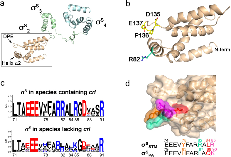
Figure 1. The Crl binding region of S. Typhimurium σS and sequence comparison of σS from species harbouring crl or lacking crl.
(a) Cartoon representation of the structural model16 of full-length σSSTM, in which domains σS2, σS3 and σS4 are shown. The helix α216,21 and the DPE motif21 within σS2 are highlighted. (b) Zoomed view of σS2 in which the side chains of critical residues are depicted in cyan and yellow. (c) WebLogo (http://weblogo.threeplusone.com/create.cgi) of σS residues 71–91 (numbered as in σSSTM) generated with the σS sequences listed in Supplementary Figure S2, from bacterial genomes containing crl or lacking crl. (d) Sequence alignment of σS helix α2 from S. Typhimurium and P. aeruginosa, both with their own numbering. Residues that differ between σSPA and σSSTM are highlighted in the alignment and represented with the same colour code as in the surface representation of S. Typhimurium σS2 shown above.

