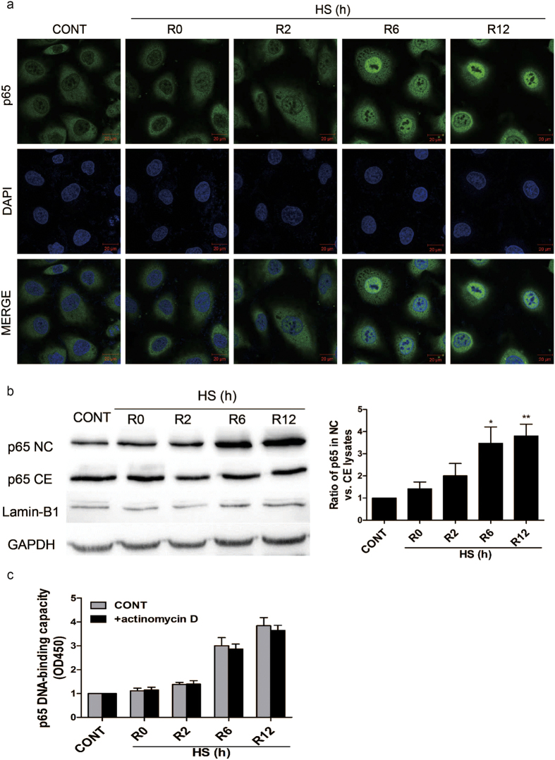
Figure 1. Relocalization of p65 from the cytosol into the nucleus of heat stressed HUVECs.
Cells were incubated at 37 °C (CONT) or were subjected to a heat stress (HS) treatment at 43 °C for 90 min, followed by a recovery period at 37 °C for 0 h (R0), 2 h (R2), 6 h (R6), or 12 h (R12). (a) The cells were then fixed and processed for indirect immunofluorescence analysis using an antibody raised against the p65 subunit of NF-κB. Representative images are shown. (b) Expression of p65 was detected in cytoplasmic (CE) and nuclear (NC) fractions of HUVECs by Western blotting. The cropped images represent blotting experiments that were performed under the same experimental conditions. (c) HUVECs were pretreated with or without 0.5 μg/ml actinomycin D 5 min before being incubated at 37 °C (CONT) or being subjected a HS treatment. Whole cell extracts were prepared and NF-κB binding to DNA was quantified with the Trans-AMTMp65 transcription factor assay kit. Each value represents the mean ± SD of three independent experiments. **P < 0.01 and ***P < 0.001 versus control.

