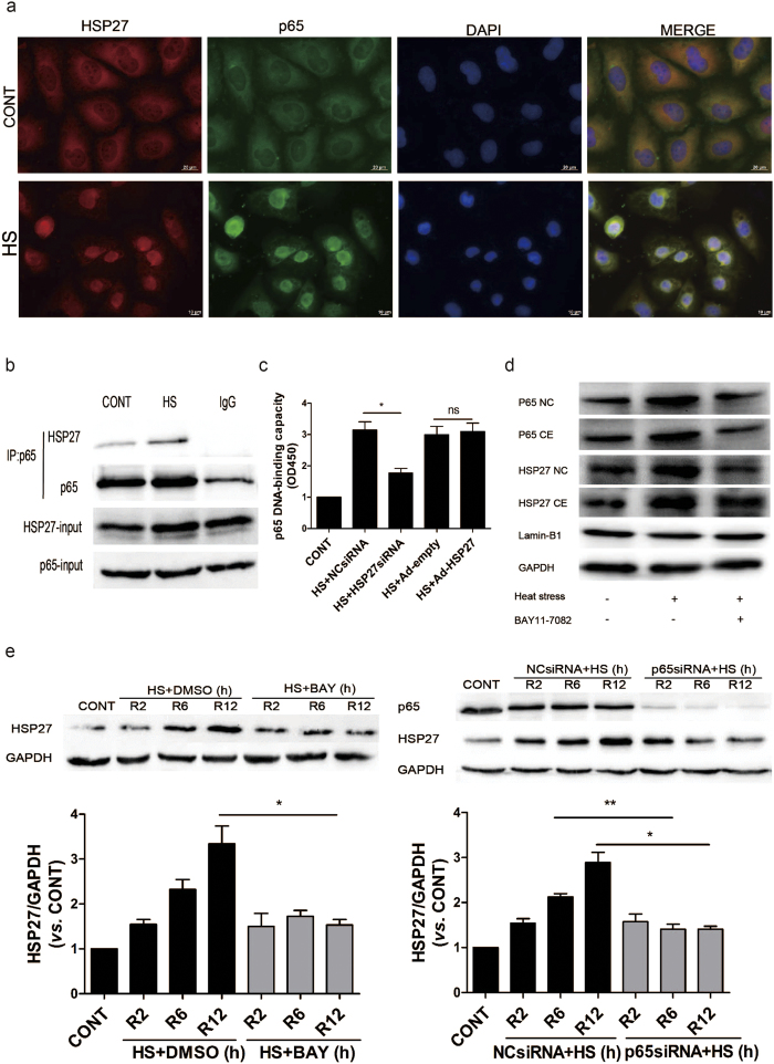
Figure 5. HSP27 associates and interacts with NF-κB in heat stressed HUVECs.
HUVECs were incubated at 37 °C (CONT) or were subjected to a heat stress (HS) treatment at 43 °C for 90 min, then were further incubated for 6 h at 37 °C. (a) The cells were subsequently stained with an anti-HSP27 antibody (shown in green) and an anti-p65 antibody (shown in red). Co-staining with DAPI was used to visualize the nuclei (shown in blue). Merged images of these three stainings are shown in the right panels. (b) Whole cell lysates were prepared and probed in co-immunoprecipitation assays with an anti-p65 antibody, while a normal IgG antibody was used as a negative control. Western blot assays were then performed for the immunoprecipitated samples with an anti-HSP27 antibody. (c) HUVECs were transfected with a negative control siRNA (NCsiRNA) or an Hsp27-targeted siRNA (HSP27siRNA), or with an empty adenovirus vector (Ad-empty) and a adenovirus vector expressing HSP27 (Ad-HSP27), respectively. These transfected cells were then incubated at 37 °C (CONT) or were subjected to a heat stress (HS) treatment at 43 °C for 90 min, and recovery period at 37 °C for 6 h. Binding of NF-κB to DNA was quantified by using a Trans-AMTMp65 transcription factor assay kit. The values represent the relative binding that was measured at 450 nm. (d–e) HUVECs were pretreated with DMSO or 5 μM BAY11-7082 for 1 h (d), or were transfected with a negative control siRNA (NCsiRNA) or a p65-targeted siRNA (p65siRNA) for 48 h (e). Then both sets of cells were incubated at 37 °C (CONT) or 43 °C for 90 min (HS), followed by recovery period at 37 °C for 2 h (R2), 6 h (R6), or 12 h (R12). Expression levels of p65 and HSP27 were detected in Western blot assays performed. The cropped images represent blotting experiments that were performed under the same experimental conditions. Each value represents the mean ± SD of three independent experiments. *P < 0.05, **P < 0.01, ns: no significance. CE: cytoplasmic; NC: nuclear.

