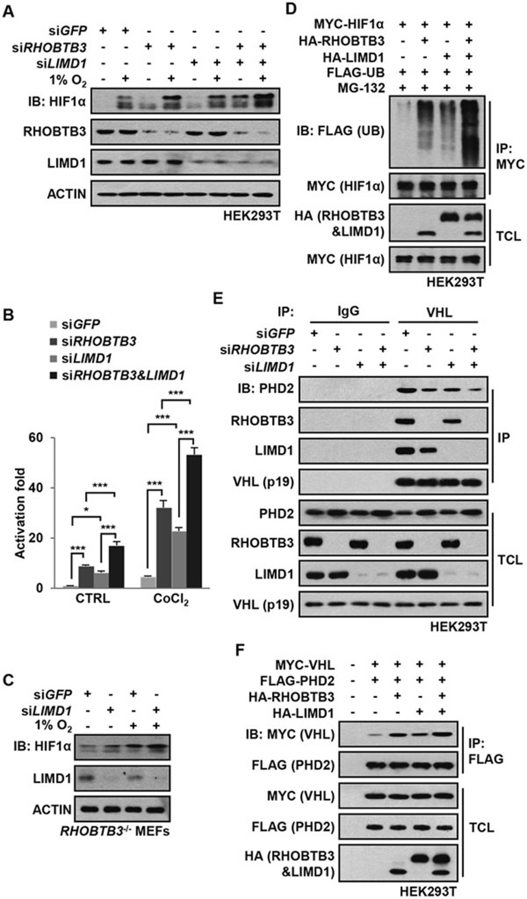
Figure 4.
RHOBTB3 and LIMD1 cooperatively regulate HIF1α. (A) RHOBTB3 and LIMD1 cooperatively suppress the protein level of HIF1α. HEK293T cells were infected with lentiviruses expressing siRNA targeting GFP, RHOBTB3 and/or LIMD1. At 16 h post-infection, cells were maintained in normoxia or exposed to hypoxia for 8 h before lysis and immunoblotting with antibodies indicated. (B) RHOBTB3 and LIMD1 cooperatively suppress the transcriptional activities of HIF1α. HEK293T cells were infected with different combinations of lentiviruses as indicated. Transcriptional activities of HIF1α were measured using a dual luciferase assay system as described in Figure 2E. Data are presented as mean ± SEM, n = 3 for each group, *P < 0.05, ***P < 0.001 (ANOVA followed by Tukey). (C) Knockdown of LIMD1 in RHOBTB3−/− MEFs further increases the protein levels of HIF1α. RHOBTB3−/− MEFs were infected with lentiviruses expressing siRNA targeting GFP or LIMD1. At 36 h post-infection, cells were maintained in normoxia or exposed to hypoxia for 8 h, before the western blot analysis. (D) RHOBTB3 and LIMD1 cooperatively promote the ubiquitination of HIF1α. HEK293T cells were transfected with different combinations of MYC-HIF1α, HA-RHOBTB3, HA-LIMD1 and FLAG-UB (ubiquitin). After treatment with 10 μM MG-132 for 10 h, the cells were lysed, and the lysates were subjected to IP with antibody against MYC (for HIF1α). The IP product was analyzed by western blotting to determine the ubiquitination levels of HIF1α. (E) Knockdown of RHOBTB3 and/or LIMD1 decreases PHD2-VHL interaction. HEK293T cells were infected with lentiviruses expressing siRNA targeting GFP, RHOBTB3 and/or LIMD1. At 16 h post-infection, cells were lysed and the endogenous VHL was immunoprecipitated, and the IP product was analyzed by immunoblotting. (F) Ectopically expressed RHOBTB3 and LIMD1 cooperatively promote PHD2-VHL interaction. HEK293T cells were transfected with different combinations of MYC-VHL, HA-RHOBTB3, HA-LIMD1 and FLAG-PHD2. Protein extracts from the transfected cells were subjected to IP with antibody against FLAG and analyzed by immunoblotting with antibodies indicated.

