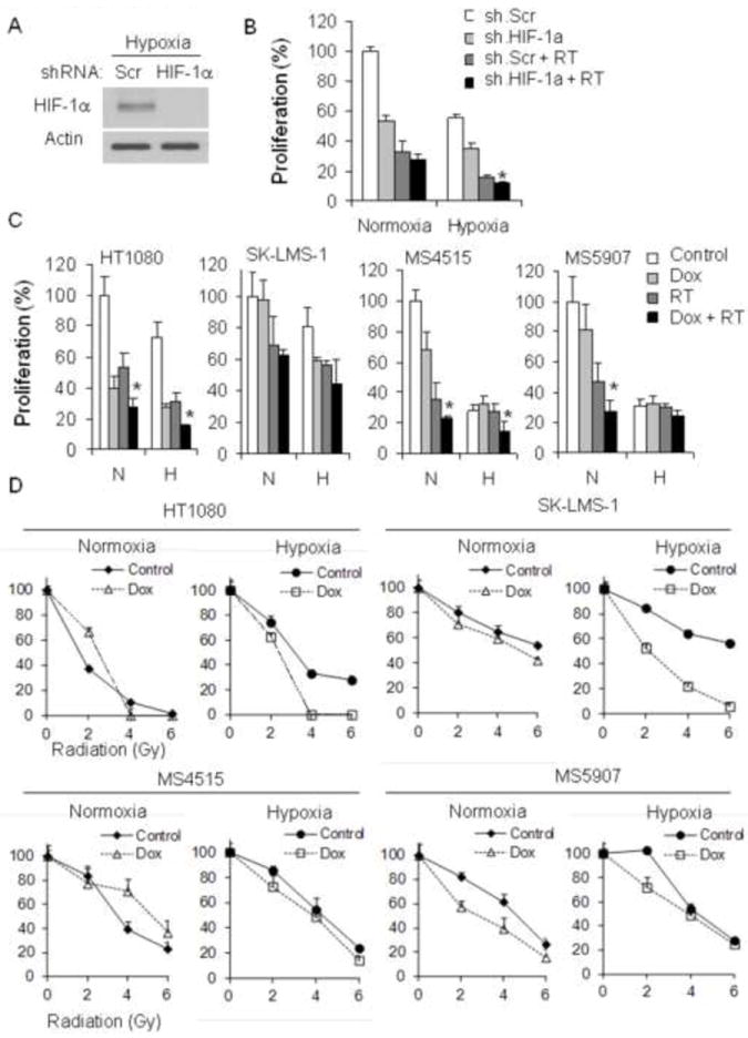
Figure 5.

(A) Western blot analysis of HIF-1α in HT1080 cells transduced with HIF-1α shRNA (sh.HIF-1α) or scrambled (Scr) shRNA. Actin blot serves as loading control. (B) Proliferation of HT1080 cells after RT (6 Gy) and/or transduction with HIF-1α shRNA (sh.HIF-1a) or scrambled shRNA (sh.Scr). (C) Proliferation of four STS cell lines 3 days after RT and/or low dose doxorubicin (0.005 μM). (D) Colony formation of four STS cell lines in normoxia and hypoxia after RT (0, 2, 4 and 6 Gy), and/or low dose doxorubicin (0.005 μM). All experiments were performed in normoxia and hypoxia. *p<0.05 compared to all other groups.
