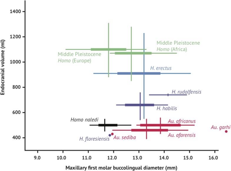
Figure 12. Brain size and tooth size in hominins.
The buccolingual breadth of the first maxillary molar is shown here in comparison to endocranial volume for many hominin species. H. naledi occupies a position with relatively small molar size (comparable to later Homo) and relatively small endocranial volume (comparable to australopiths). The range of variation within the Dinaledi sample is also fairly small, in particular in comparison to the extensive range of variation within the H. erectus sensu lato. Vertical lines represent the range of endocranial volume estimates known for each taxon; each vertical line meets the horizontal line representing M1 BL diameter at the mean for each taxon. Ranges are illustrated here instead of data points because the ranges of endocranial volume in several species are established by specimens that do not preserve first maxillary molars.

