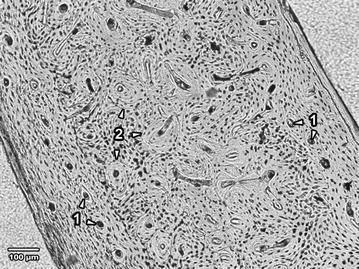
Fig. 1.

Microscopical structure of compact bone in rabbits from the control groups. 1 Primary vascular longitudinal bone tissue near endosteal and periosteal surfaces (arrows vascular canals of primary osteons). 2 Dense Haversian bone tissue creating the middle part of the substantia compacta (arrows secondary osteons)
