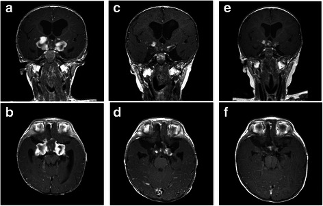
Fig. 2.

Serial MRI scans showing response to high doses chemotherapy in a five-month-old infant with hypothalamic anaplastic astrocytoma. Axial and coronal Gd-enhanced T1-weighted MR scans: immediately post-biopsy image of the hypothalamic lesion (a-b); c post-chemotherapy MRI scans demonstrating a partial response (c-d); MRI scans showing a stable disease 27 months from diagnosis (e-f)
