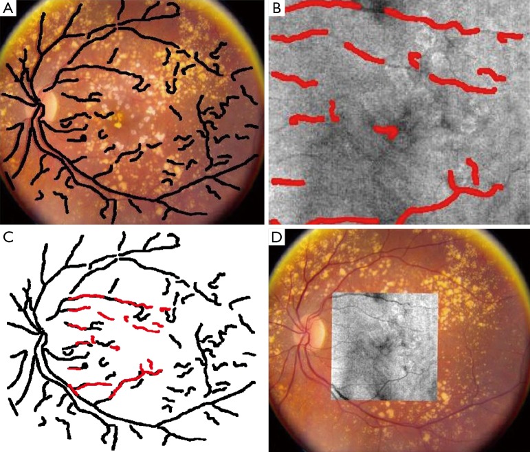
Figure 7.
Color fundus reference image superimposed by the corresponding blood vessel ridge image (A), en face OCT image superimposed by the corresponding blood vessel ridge image (B), the registration result of blood vessel ridge image (C) and intensity images (D) [(Reprinted with permission) (75)].

