
Fig. 3.
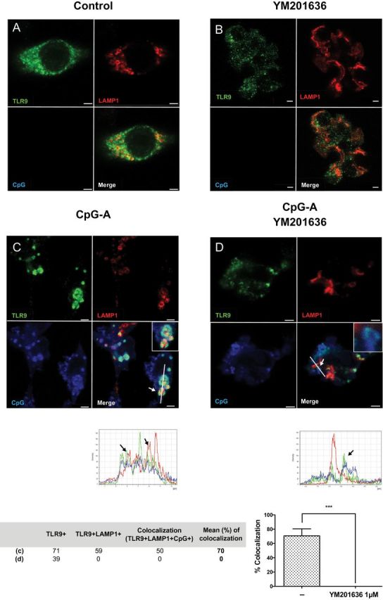
PIKfyve inhibitor blocks TLR9 localization to the LAMP1+ compartment upon CpG stimulation. RAW 264.7 cells stably transduced with TLR9-HA were stimulated with DOTAP-CpG-A-Cy5 for 6 h in the absence (C) or presence (D) of YM201636 (1 μM) given 1 h prior to CpG-A stimulation. Subsequently, cells were stained and analyzed by confocal microscopy to detect TLR9-HA (green), LAMP1 (red) and CpG (blue). Colocalization of TLR9 and LAMP1 or TLR9, LAMP1 and CpG was defined by structures in which TLR9/CpG signals were surrounded by LAMP1 staining. Scale bars, 2.5 μm. No-stimulation controls in the absence or presence of inhibitor are shown as (A), (B), respectively. ***P < 0.001.