Fig. 3.

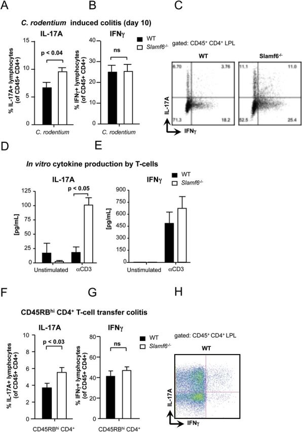
Enhanced Th17 response in Slamf6 −/− mice during C. rodentium-induced colitis. WT and Slamf6 −/− mice were infected by oral gavage of 2×10⁹ C. rodentium bacteria. At day 10 post-infection, leukocytes were isolated from the colon lamina propria. The percentage of (A) IL-17A+ and (B) IFNγ+ lymphocytes is represented as a percentage of total CD45+ CD4+ lymphocytes. (C) Representative dot plots of intracellular staining for IL-17A and IFNγ obtained from WT and Slamf6 −/− mice. Isolated CD4+ splenocytes were cultured o/n in the presence of plate-bound anti-CD3 antibody. The amount of (D) IL-17A and (E) IFNγ cytokines in the supernatant of these cultures is represented. WT and Slamf6 −/− CD45RBhi CD4+ splenocytes were transferred into Rag −/− mice. The percentage of (F) IL-17A+ and (G) IFNγ+ lymphocytes is represented as a percentage of total CD45+ CD4+ lymphocytes. (H) Representative dot plot of intracellular staining for IL-17A and IFNγ obtained from Rag −/− mice in which WT T cells were transferred.
