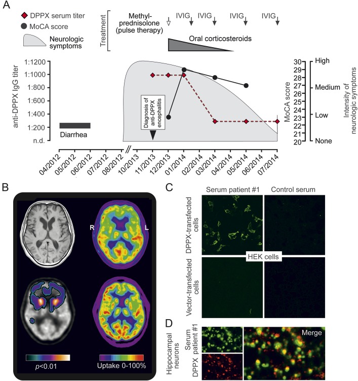
Figure 1. Clinical and paraclinical findings of a novel patient with anti–dipeptidyl-peptidase-like protein 6 encephalitis.
(A) Time course of the disease, intensity of neurologic symptoms, Montreal Cognitive Assessment (MoCA) test scores, anti–dipeptidyl-peptidase-like protein 6 (DPPX) antibody titers, and treatments of a newly identified patient (patient 1) with anti-DPPX encephalitis. (B) T1-weighted axial MRI demonstrates no significant atrophy (upper left). PET with F-18-fluorodeoxyglucose (FDG-PET) as marker of synaptic activity shows strongly reduced activity bilaterally in the heads of the caudate nuclei and mild to moderate reduction in the frontal cortex (upper right); compared with FDG-PET of a healthy person (lower right). Reduction of activity in the heads of the caudate nuclei and frontal cortex was confirmed by voxel-based testing vs a healthy control group, demonstrating statistical significance at the uncorrected α = 0.01 level (lower left; brighter colors indicate lower p values). (C) Binding of patient 1 serum (1:100 dilution) to DPPX-transfected HEK293 cells. Healthy control serum (1:10 dilution) and HEK293 cells transfected with empty vector served as controls. (D) Primary hippocampal neurons were incubated for 2 hours with patient 1 serum (1:100 dilution), fixed, and double-stained for human immunoglobulin G (IgG) and DPPX using a commercial anti-DPPX antibody. The merge demonstrates overlap of both signals (confocal images). IVIG = IV immunoglobulin; n.d. = not detectable.

