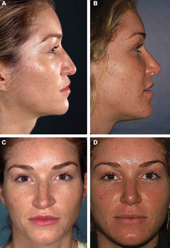
Fig. 2.

Fat grafting and rhinoplasty. Preoperative lateral (A), postoperative lateral (B), preoperative frontal (C), and postoperative frontal (D) images of a 27-year-old white woman who underwent autologous fat grafting to the nasofrontal and pyriform regions in combination with open rhinoplasty. The preoperative photographs demonstrate a “premature” forehead, atrophy of the glabella and radix, and an acute nasolabial angle. The postoperative photographs, obtained 6.5 years after the procedures, show improved contour of the lower forehead after transplantation of 19 cm3 of fat to the nasofrontal region and 16.5 cm3 to the pyriform region. Note reduction of the nasofrontal angle (from 133.4° preoperatively to 124.6° postoperatively) and greater tip rotation, with significant change in the nasolabial angle (from 51.3° preoperatively to 85.8° postoperatively).
