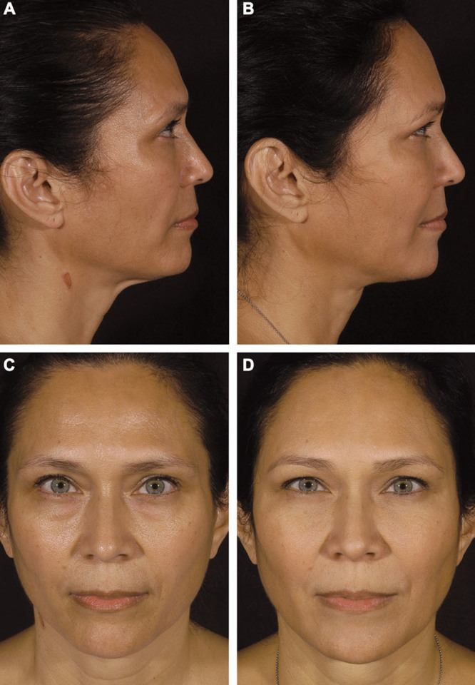
Fig. 5.

Fat grafting alone. Preoperative lateral (A), postoperative lateral (B), preoperative frontal (C), and postoperative frontal (D) images of a 51-year-old white woman who underwent autologous fat grafting of the face. The preoperative photographs demonstrate loss of forehead volume and contour, with an obtuse nasofrontal angle and an appropriately rotated nasal tip. The postoperative photographs, obtained 5.8 years after the procedure, show greater fullness and contour of the forehead. The patient received 19 cm3 of fat in the nasofrontal region. Note improvement in the nasofrontal angle (from 137.4° preoperatively to 130.3° postoperatively) and the slight change in the nasal tip (from 94.5° preoperatively to 96.3° postoperatively) presumably due to malar, nasolabial, and upper white lip autologous fat augmentation.
