
FIGURE 5.
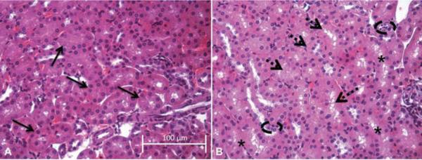
Representative hematoxylin and eosin (H&E) images showing changes in proximal tubule morphology. Images (40×) are representative of the results for n = 3 control aniumals (A) and n = 4 CdO NP-treated (B) animals at GD 10.5; scale bar indicates a length of 100 μm. Solid arrows in (A) designate individual round tubules in the control kidney section. Dashed arrows (B) denote tubules with less intense cytosolic staining, while broken ovals highlight areas of small, irregularly shaped nuclei; asterisks designate areas of gaps or vacuoles.