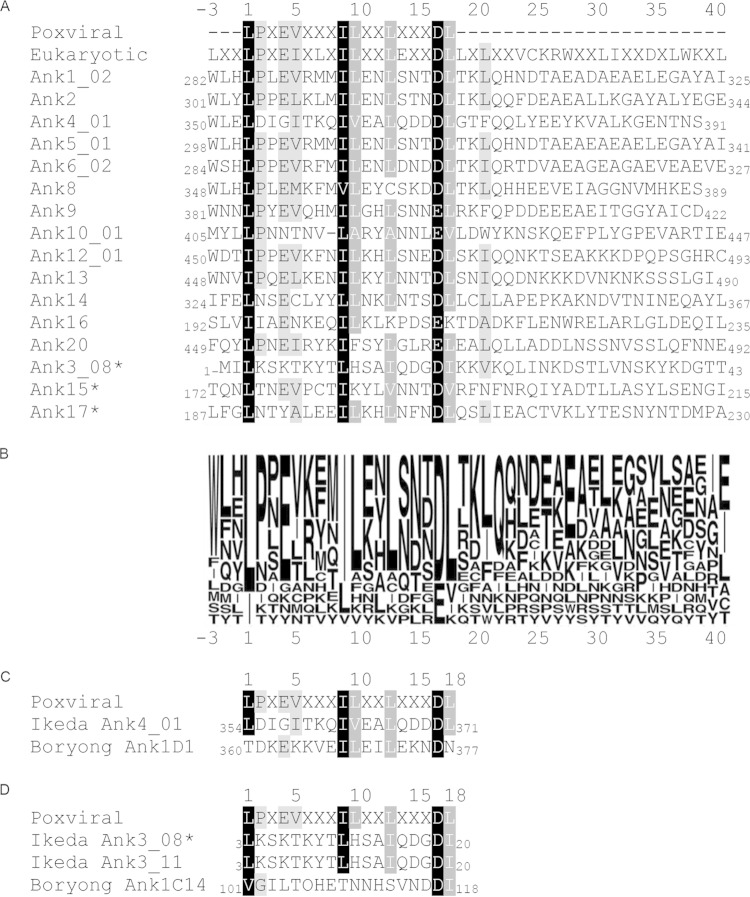
FIG 7.
Identification of highly conserved O. tsutsugamushi strain Ikeda Ank F-box residues. (A) Alignment of F-boxes from the 16 O. tsutsugamushi strain Ikeda Anks that bind SKP1 with canonical poxviral and eukaryotic F-box sequences. Regions containing each Ank protein's F-box domain were aligned to canonical poxviral and eukaryotic F-box sequences using Geneious software. The shaded residues indicate amino acid similarity, with black highlighting residues that are similar among 100% of the aligned sequences, dark gray highlighting residues that are similar among 83.3 to 94.4% of the aligned sequences, and light gray highlighting residues that are similar among 72.2 to 77.8% of the aligned sequences. Residues were considered similar if they fell within the following groups: A, V, L, I, and M; D and E; K and R; P and G; or F, Y, and W (39). The three Anks marked with asterisks contain a predicted F-box sequence but interacted weakly with SKP1 in pulldown assays compared to the other 13 Anks, as shown in Fig. 2. The numbers at the N and C termini of each sequence indicate the amino acid positions of the F-box motif within the full-length Ank. (B) Sequence logo representation corresponding to the alignment in panel A, showing Ank amino acid conservation at each position. The letter size indicates the relative frequency of a given residue at its respective position in the alignment. (C) Alignment of the Ikeda Ank4_01 F-box sequence with that of its Boryong homolog, Ank1D1. (D) Alignment of the sequence of the Ikeda Ank3_08 F-box, which was shown to bind weakly to SKP1 in this study (Fig. 2), with that of its Ikeda paralog, Ank3_11, and its homolog, Boryong Ank1C14. The numbers above (A, C, and D) and below (B) the alignment serve as references for amino acid positions. The number 1 denotes the first position of the canonical poxviral F-box residue.

