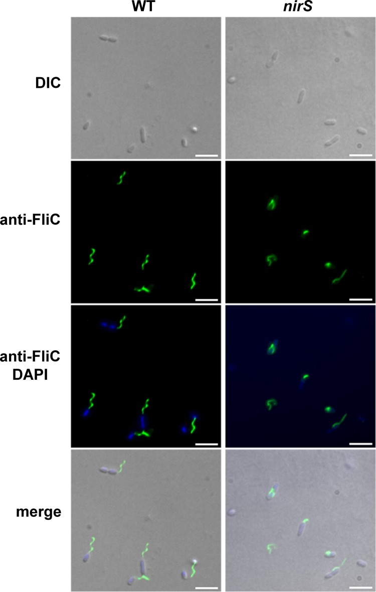
FIG 3.
Flagellar morphology in the wild type and nirS mutant visualized by immunofluorescence microscopy. For visualization of extracellular FliC protein, samples of bacteria cultured anaerobically in LB containing arginine were labeled with primary rabbit anti-FliC antibody followed by secondary Alexa Fluor 488-conjugated goat anti-rabbit antibody (green), and the DNA was labeled with DAPI (blue). Arrowheads point to nonfunctional flagella in the NirS mutant strain. Fluorescence images were taken and processed as described in Materials and Methods. Scale bars, 10 μm.

