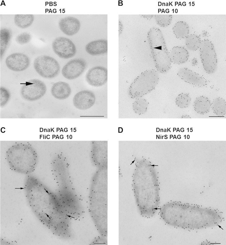
FIG 4.
Cellular localization and colocalization of DnaK, FliC, and NirS in anaerobically grown, denitrifying cells of P. aeruginosa PA14. Cells were prepared for transmission electron microscopy, treated with specific antibody, and protein A/G conjugated with 10- or 15-nm-diameter gold nanoparticles (PAG 10 and PAG 15, respectively), counterstained, and examined in a TEM910 transmission electron microscope, as described in Materials and Methods. (A) Antibody-negative control with PAG 15; (B) anti-DnaK antibodies with PAG 15, followed by protein A blocking, and then PAG 10; (C) anti-DnaK antibodies with PAG 15, followed by protein A blocking, and then anti-FliC antibodies with PAG 10; (D) anti-DnaK antibodies with PAG 15, followed by protein A blocking, and then anti-NirS antibodies with PAG 10. DnaK is seen to be distributed mostly in the extracytoplasmic region; several cocomplexes between DnaK and FliC as well as DnaK and NirS are observed and are indicated by arrows. Scale bars are 500 nm (A and B) and 200 nm (C and D).

