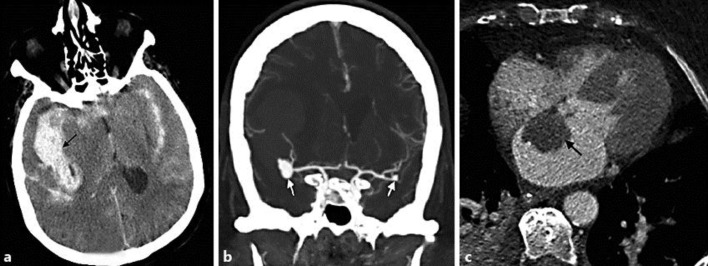
Fig. 1.
a Noncontrast CT of the head demonstrates diffuse subarachnoid hemorrhage with a clot in the right sylvian fissure region. b CT angiogram of the brain shows a 9-mm lobulated aneurysm at the right MCA trifurcation and a small 3-mm aneurysm at the left MCA bifurcation. c Axial high-resolution CT of the heart shows a left atrial myxoma measuring 3.2 × 2.3 cm.

