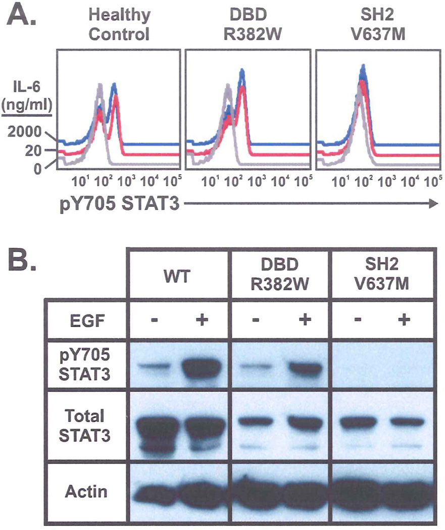
Figure 3.
A. Phospho-flow cytometry showing IL-6 induced tyrosine phosphorylation of STAT3 is similar in control and DBD (R382W) mutant PBMCs but decreased in SH2 (V637M) mutant PBMCs. The defect is not overcome with 100x IL-6 concentration. B. EGF induced tyrosine phosphorylation of GFP-STAT3 in transiently transfected Cos-7 cells. Lower band in total STAT3 lanes is a degradation product of transfected STAT3 (data not shown).

