
Figure 4.
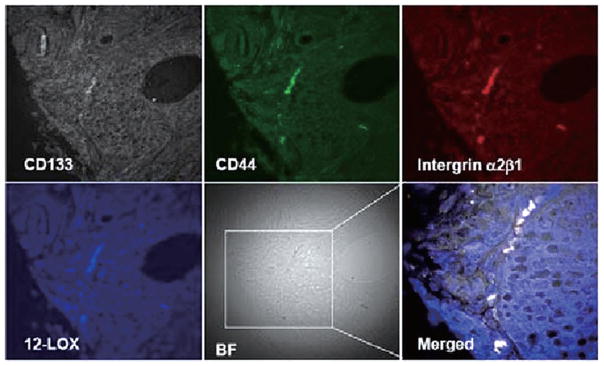
PCa SCs were identified by the markers of CD44+/integrin α2β1high/CD133+ in PCa tissue samples and overexpression of 12-LOX (blue) was shown by using 4-channel confocal microscopy (40×10). BF, bright field.
Official websites use .gov
A
.gov website belongs to an official
government organization in the United States.
Secure .gov websites use HTTPS
A lock (
) or https:// means you've safely
connected to the .gov website. Share sensitive
information only on official, secure websites.
PCa SCs were identified by the markers of CD44+/integrin α2β1high/CD133+ in PCa tissue samples and overexpression of 12-LOX (blue) was shown by using 4-channel confocal microscopy (40×10). BF, bright field.