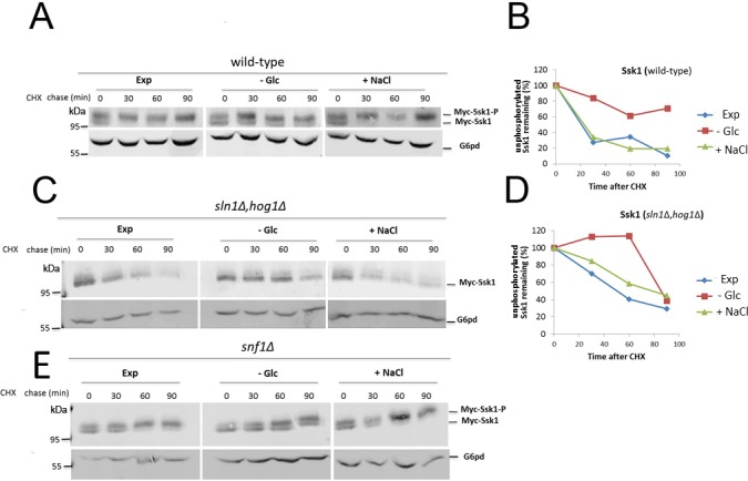
Fig 3. Ssk1 is stabilized during glucose starvation.

Wild-type (A) and sln1Δhog1Δ (C) cells expressing Myc-Ssk1 were exponentially grown in HC-Leu (EXP), and then starved for glucose (-Glc) or subjected to osmotic stress (+NaCl). Cycloheximide was added to stop protein synthesis and cells were collected at the indicated times. Levels of unphosphorylated Ssk1 were monitored using SDS-PAGE followed by immunoblotting with anti-Myc antibody. Glucose-6-phosphate dehydrogenase (G6pd) was detected as the loading control. The relative quantities of unphosphorylated Ssk1 in wild-type (B) and sln1Δhog1Δ cells (D) were normalized using G6pd levels. E—snf1Δ cells expressing Myc-Ssk1 were exponentially grown in HC-Leu (EXP), and then starved for glucose (-Glc) or subjected to osmotic stress (+NaCl). Cycloheximide was added to stop protein synthesis and cells were collected at the indicated times. Levels of unphosphorylated Ssk1 were monitored using SDS-PAGE followed by immunoblotting with anti-Myc antibody. Glucose-6-phosphate dehydrogenase (G6pd) was detected as the loading control.
