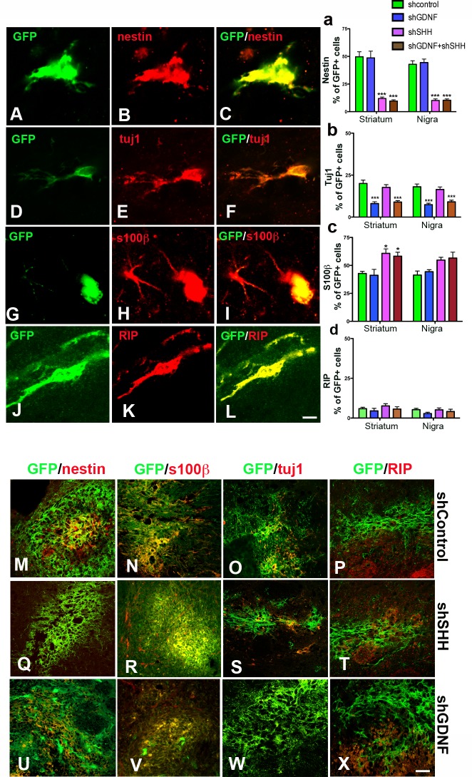
Fig 5. Inhibition of SHH influences the phenotypic fate of grafted NPCs.
We analyzed the co-expression of antigens specific to nestin (undifferentiated NPCs, A-C), immature neurons (tuj1, D-F), astrocytes [(s100β (G-I) and oligodendroglia [RIP (J-L)] in the GFP NPCs. Counts of grafted GFP cells indicated that while the number of nestin+ cells was significantly lower, compared to control NPCs in donor cells silenced for SHH or GDNF and SHH, shGDNF grafts did not show such an effect (a). This was true for grafts both in the striatum as well as SN. Representative images of nestin stained grafts are shown in M, Q, U. The GDNF silenced grafts on the other hand showed a significant (p<0.001) reduction in the number of cells expressing the neuron marker tuj1 (b). Such a decline in tuj1 expression was not observed in the SHH silenced NPCs. Representative images of tuj1 stained grafts are shown in O, S, W. Further, the number of S100β+ cells was greater in striatal grafts with SHH knock-down, with no such influence observed in any of the other experimental groups (c). Representative images of S100β+ cells in grafts are shown in N, R, V. Finally, with respect to oligodendrocyte differentiation, no significant differences were observed between control and silenced NPCs (d). Representative images of RIP staining in grafts are shown in P, T, X. Values are expressed as mean ± SEM [*p<0.05, **p<0.01, ***p<0.001 compared to shC, one way ANOVA with Bonferroni’s post test]. Scale bars: A-L—10 μm; M-X—30 μm.

