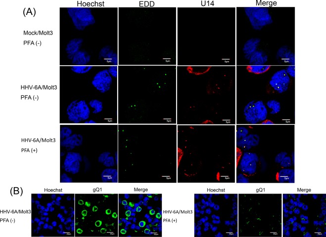
Fig 2. Subcellular localization of U14 and EDD in HHV-6A-infected cells.
HHV-6A- or mock-infected Molt3 cells, with or without PFA, were harvested at 24 h post-infection (pi), fixed, and subjected to IFA using antibodies against U14 and EDD (A) or gQ1(B); nuclear DNA was counterstained with Hoechst 33342. Merged panels show colocalized U14 and EDD in nuclei (A). gQ1 was detected in the cells without PFA, but not with PFA (B). Scale bars: 5 μm (A) and 20μm (B). Single sections are shown.

