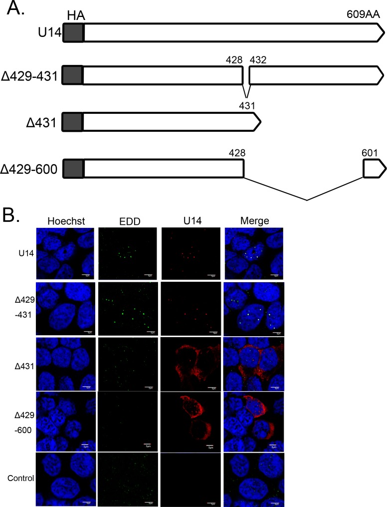
Fig 4. Schematic diagram of HHV-6A U14, its deletion mutants, and the localization of each mutant.
(A) The HHV-6A strain U1102 U14 and its deletion mutants (white box) were N-terminally fused to the HA tag (gray box). Numbers indicate positions in the amino acid sequence of U14. (B) 293T cells were transfected with HHV-6A U14, its deletion mutants, or empty vector as a negative control. The cells were harvested at 12 h post-transfection, fixed, and subjected to IFA using antibodies against HA (for U14) and EDD; nuclear DNA was counterstained with Hoechst 33342. Scale bars: 5 μm. Single sections are shown.

