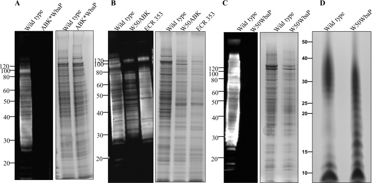
Fig 3. Absence of A-LPS in W50ABK*WbaP and W50WbaP mutants.
Immunoblot analysis of whole cell culture lysates with anti-APS (mAb 1B5) antibody. (A) WT and W50ABK*WbaP mutant, (B) WT, W50ABK mutant original stock and ECR353 (another ABK mutant in W50), (C) WT and W50WbaP mutant. Images on the right of each immunoblot (A-C) shows the corresponding commassie stained gel to show a loading control. (D) Silver stained LPS profiles from WT and W50WbaP.

