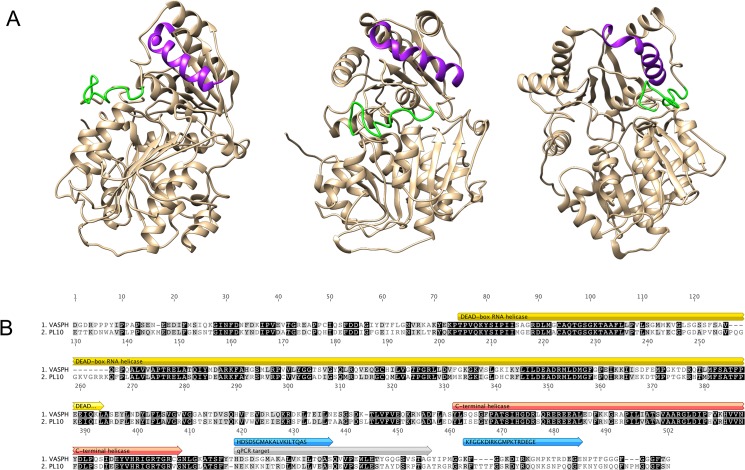
Fig 4. VASA homolog of Ruditapes philippinarum (VASPH).
(A) VASPH structure model. Chimera 1.8.1 was used to model the protein structure. Peptides used for antibody production are highlighted: HDS in purple; KFG in green. (B) Alignment of VASPH and PL10 of R. philippinarum. Peptide location (blue), qPCR target (grey) and protein main domains (yellow and red) are highlighted.

