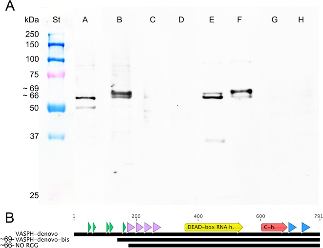
Fig 5. Detection of VASPH variants.
(A) Anti-VASPH specificity: Western blots of ovary extracts (Oe) and testis extracts (Te) of adults. From left to right: St: protein standard. A: Oe/anti-VASPH-HDS. B: Te/anti-VASPH-HDS. C: Oe/anti-VASPH-HDS control. D: Te/anti-VASPH-HDS control. E: Oe/anti-VASPH-KFG. F: Te/anti-VASPH-KFG. G: Oe/anti-VASPH-KFG control. H: Te/anti-VASPH-KFG control. Western blots were obtained with the loading of 15 μg of homogenate per lane. (B) Different VASPH transcript assemblies highlighted different N-terminus of the protein. RGG motifs (green), zinc fingers (violet), protein main domains (yellow and red), and peptide location (blue) are highlighted.

