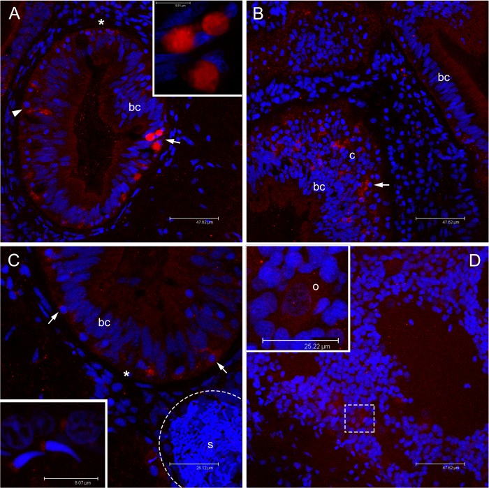
Fig 6. Immunolocalization of VASPH in germ cells of juveniles approaching their first reproductive season.
(A) In juvenile clams, anti-VASPH highlighted many immunospots in germ cells with rounded nucleus (PGCs) localized in the thickness of intestinal epithelium, between unstained batiprismatic cells (bc) of the gut, and the basal lamina (asterisk). In some germ cells, the immunospots appear separated (arrowhead), while in other cells the spots aggregate at a side of the cell cytoplasm (arrow; magnification in the inset). Scale bar = 47.62 μm (inset scale bar = 8.61 μm). (B) Stained germ cells (arrow) are also visible in the connective tissue (c) between two intestinal loops. Scale bar = 47.62 μm. (C) Portion of young male section that shows an acinus (dashed circle) full of sperm (s) close to an intestinal loop in which some stained germ cells are present (arrows). The inset shows a magnification of two spermatozoa with a lightly stained mitochondrial midpiece and a spermatid (up right) with a big immunostained spot near the nucleus. Scale bar = 26.12 μm (inset scale bar = 8.07 μm). (D) Portion of young female section that shows early oocytes at different stage of development with a light VASPH staining in the cytoplasm. In the inset, a magnification of an early oocyte (o) showing few small granules. Scale bar = 47.62 μm (inset scale bar = 25.22 μm). Red: VASPH staining; blue: nuclear staining.

