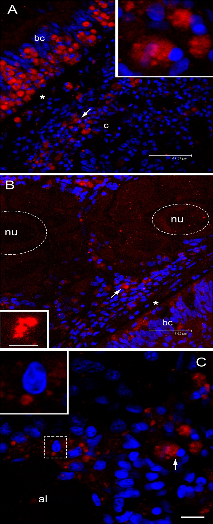
Fig 7. Immunolocalization of VASPH in germ cells of gametogenic adult females.

(A) Section with a portion of intestine and connective tissue. Inside the intestinal epithelium, among batiprismatic cells (bc), a strong proliferation of VASPH stained germ cells (arrow) is observed. Many germ cells (arrow; magnified in the inset) have passed the basal lamina (asterisk) to the connective tissue (c). Scale bar = 47.57 μm. (B) In the connective tissue, in proximity of the intestine, germ cells (arrow) surround acini full of eggs (two eggs are highlighted with a dashed oval; nu: nucleus). In the egg, small stained granules are scattered in the cytoplasm (inset: granule magnification). Scale bar = 47.62 μm (inset scale bar = 4.87 μm). (C) At the periphery of an acinus lumen (al), very early oocytes of about 10 μm show big stained spots (one oocyte is magnified in the inset). Scale bar = 10.54 μm. Red: VASPH staining; blue: nuclear staining.
