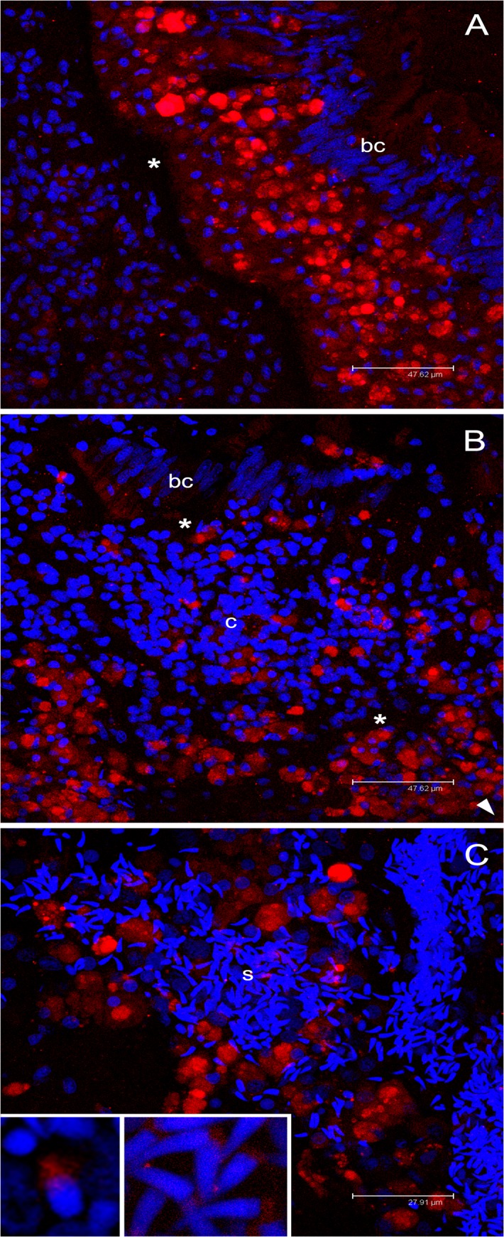
Fig 8. Immunolocalization of VASPH in germ cells of gametogenic adult males.

(A) Strong proliferation of VASPH-stained germ cells in the intestinal epithelium at one side of the basal lamina (asterisk). Batiprismatic cells (bc) resulted VASPH-unstained. Scale bar = 47.62 μm. (B) Many stained germ cells in the connective tissue (c) between two intestinal loops (the arrowhead points to the position of other batiprismatic cells of the gut). Scale bar = 47.62 μm. (C) High magnification of a portion of male acinus showing many spermatozoa that fill the lumen. Inset on the left: spermatid with VASPH staining limited at the posterior part of the elongating nucleus. Inset on the right: several spermatozoa showing an even more reduced labelling in the mitochondrial midpiece. Scale bar = 27.91 μm. Red: VASPH staining; blue: nuclear staining.
