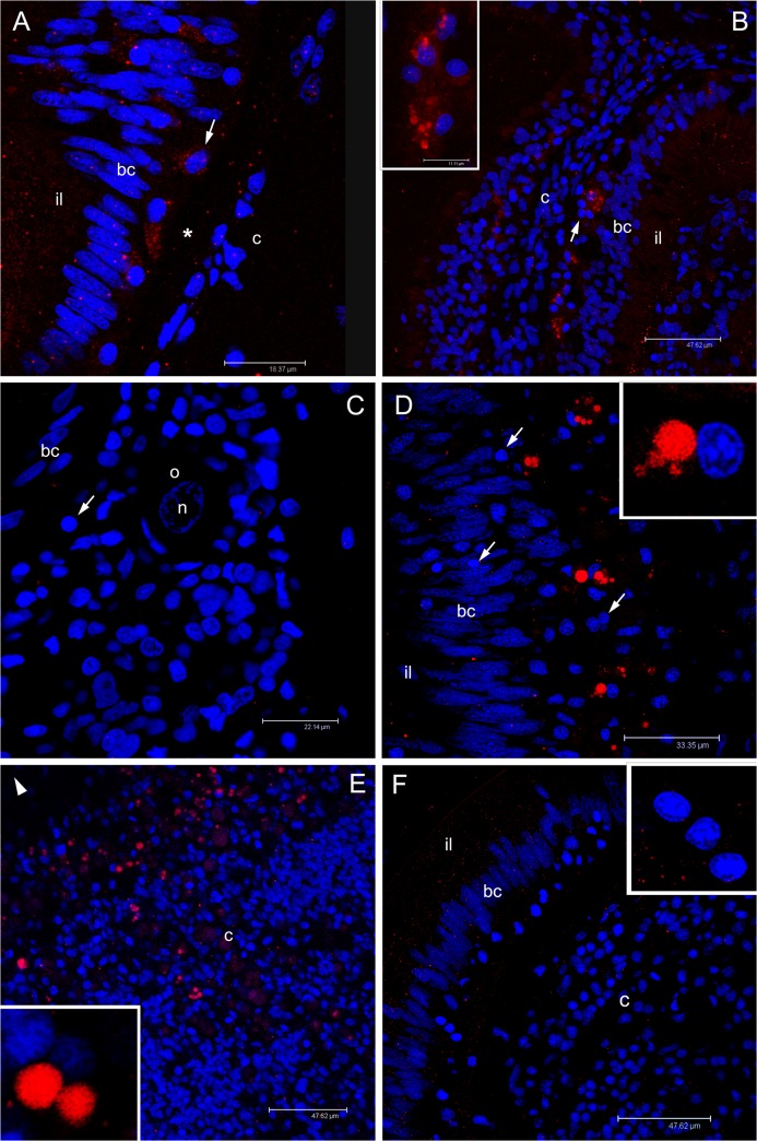
Fig 9. Immunolocalization of RPHM21 in germ cells.
(A) In juveniles, the immunological reaction highlighted some rounded-nucleus cells (germ cells) with a diffused cytoplasmic RPHM21 staining (arrow) between unstained batiprismatic cells (bc) and the basal lamina (asterisk). Intestinal lumen = il; connective tissue = c. Scale bar = 18.37 μm. (B) In some sections of juvenile animals, germ cells are visible in the connective tissue (c) and show big stained spots (inset). Scale bar = 47.62 μm (inset scale bar = 11.11 μm). (C) In some female sections, simple acini, at the beginning of their organization, sometimes containing a single oocyte (o), were found. In these female sections, germ cells were visible (arrow) but no RPHM21 staining was present. n = oocyte nucleus. Scale bar = 22.14 μm. (D) Adult male section that shows RPHM21 stained germ cells close to batiprismatic cells (bc); some germ cells do not show any RPHM21 staining (arrow). Scale bar = 33.35 μm. (E) In male connective tissue, several RPHM21-stained germ cells are found (the arrowhead points to intestine position). The inset shows magnified RPHM21stained germ cells. Scale bar = 47.62 μm. (F) In adult female sections, no RPHM21-staining was detected in germ cells (magnified in the inset). Scale bar = 47.62 μm. Red: RPHM21 staining; blue: nuclear staining.

