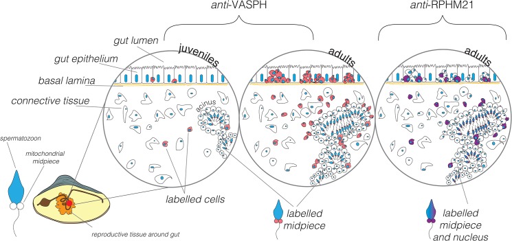
Fig 10. Scheme of RPHM21 and VASPH immunolocalization in germ cells during male gonad formation.
VASPH expression (in red): in juvenile males (first circle on the left) few, stained PGCs are localized in the intestine among batiprismatic cells, and other stained germ cells are found in the connective tissue or around the few simple-structured acini localized in the connective tissue. In gametogenic males (circle in the middle) PGCs are massively proliferating among batiprismatic cells and are strongly immunostained. In mature male acini full of spermatozoa, a diffused VASPH-staining is present in the spermatogenic cells located near the acinus wall (see also [10]). Spermatozoon midpiece appears slightly stained. RPHM21 expression (in violet): only a subpopulation of PGCs located in the intestinal epithelium appears to express RPHM21, other PGCs, recognizable for their round nucleus, result completely negative to the RPHM21 staining. Some cells with a weak RPHM21 labelling (spermatogenic cells) are recognizable in the acinus wall [41]. RPHM21 is expressed in mature spermatozoa localized in the acinus lumen, both in mitochondria and the nucleus [41]. The staining of both factors (VASPH and RPHM21) is almost always condensed in a big cluster at one side of the cell cytoplasm.

