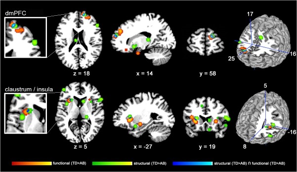
Fig 3. Structural and functional neuroimaging findings in youths with AB co-localize in right dorsomedial prefrontal cortex (dmPFC) and left insular cortex.

2-D slices displaying the thresholded and binarized ALE maps of significant overlap (P<0.05, FDR-corrected) in studies of structural (green) and functional (red) alterations in adolescents with AB (TD>AB) as well as a conjunction analysis (blue) overlaid on the Colin T1-template in MNI space. The upper-row including left cut-out as well as right surface-model highlight the right dmPFC where structural and functional alterations co-localize. The lower-row including left cut-out as well as right surface-model illustrate left insular cortex/claustrum where structural and functional alterations overlap.
