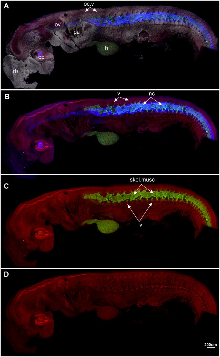
Fig 3. Callorhinchus milii, stage 23.
A-D, lateral view showing A, staining as in Fig 2, also DAPI (cell nuclei, white) staining. Blue colour indicates collagen type II staining in the notochord. B, As in A, but DAPI staining not visualised; C, Sox9 and Mf20 staining, showing dorsal and ventral vertebral elements (neural and haemal arches) and skeletal musculature; D, Sox9 staining alone. All vertebral elements separate and distinct at this stage. Abbreviations as in Fig 1, also: nc, notochord; oc.v, occipital vertebrae; skel.musc, skeletal musculature; rb, rostral bulb [28].

