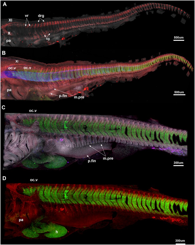
Fig 4. A-E, Callorhinchus milii, stages 25 (A, B), 27 (C, D).
A, Cell nuclei (white). Sox9 (red) marks dorsal and ventral vertebral elements and neural crest cells; the vagus and accessory nerves (X and XI) are clearly visible. Three anteriormost occipital vertebrae (oc.v) lack the spinal nerves associated with more posterior vertebrae; they appear closer together differ in shape relative to the more posterior vertebrae, which are better developed and still distinct from each other. B, notochord (blue) and developing skeletal musculature are visualized. The pectoral fin bud is beginning to develop (p.fin), while the pectoral fin musculature is differentiating from the ventral myotome (m.pre). C, Stage 27, Sox9 no longer stains the vertebral elements (differentiated beyond the prechondrogenic cartilage stage); they are best seen via DAPI staining (cell nuclei). All vertebral elements are still separate from one another. Developing pectoral fin musculature is more distinctly bifid in shape. Abbreviations as in previous figures, also: drg, dorsal root ganglia; m.pre, pectoral fin muscle precursor; p.fin, pectoral fin; sg, sympathetic ganglia; vr, ventral root.

