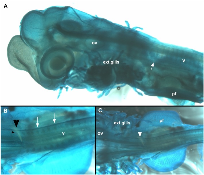
Fig 5. Callorhinchus milii, stage 28, A-C.
Cleared and stained specimen (Alcian blue). A, lateral view, showing embryo with external gills and developing vertebrae. White arrow indicates anterior vertebrae that have become misshapen, indicating fusion and coalescence. The position of these relative to the pharyngeal arches and pectoral fin indicates that these are fusing to the occipital region of the braincase, rather than as part of a more posterior synarcual. More posterior vertebrae appear normal at this stage. B, C, dorsal view, arrowhead indicates occipital anteriorly and vertebrae posteriorly. White arrow indicates foramina Abbreviations, as in previous figures, also: ext.gills, external gills.

