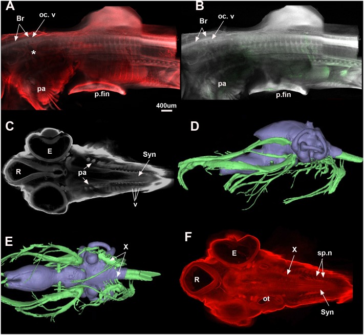
Fig 6. Callorhinchus milii.
A, B, Stage 29 (maximum projection, Sox9 red, DAPI white, Mf20 green) shows that the occipital vertebrae are still distinct from each other but that the braincase is now well developed. The posterior elements are still separate but show distinct dorsal and ventral elements for each vertebra. In stage 30 (C, F; one plane in Z, Sox9 red, DAPI white), the synarcual has started to form and can be recognised medially. The individual vertebral elements are still visible laterally (v). The vagus and accessory nerves are visible lateral to the mineralised braincase (C). D, E show distribution of the cranial nerves in a C. milii hatchling (Khonsari et al. 2013). Abbreviations as in previous figures, also Br, Braincase; E, Eye; R, Rostrum; Syn, synarcual. Images in Fig 6D and 6E taken as screenshots from a 3-D reconstructed CT scan model; Wikimedia Commons datafile, Khonsari et al. 2013. BMC Biology. doi:10.1186/1741-7007-11-27.

