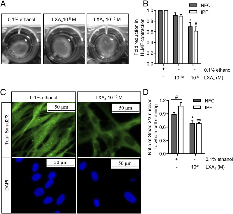
FIGURE 3.
Basal HLMF contraction and Smad2/3 nuclear translocation are inhibited by LXA4. (A) Representative images from an NFC donor showing the spontaneous contraction of HLMFs within a collagen gel and its suppression by LXA4. (B) Constitutive HLMF contraction was significantly attenuated by LXA4 (10−8 mol) in both NFC- (n = 6) and IPF-derived (n = 5) cells (two-way ANOVA corrected by Sidak multiple-comparison test; NFC, *p = 0.0009; IPF, #p = 0.0001). (C) Representative images from an IPF donor displaying total Smad2/3 expression within HLMFs. At baseline, the HLMFs have staining both in the nucleus and within the cytoplasm, which is reduced upon incubation with LXA4 (10−8 mol) for 1 h. (D) The ratio of total Smad2/3 nuclear to whole-cell staining was assessed by measuring grayscale intensity. IPF-derived (n = 6) HLMFs demonstrated increased expression of total Smad2/3 within the nucleus in comparison with NFC donors (n = 6) (#p = 0.043, Mann–Whitney U test). LXA4 (10−8 mol) significantly attenuated the amount of Smad2/3 localized to the nucleus in both NFC- and IPF-derived cells (two-way ANOVA corrected by Sidak multiple-comparison test; NFC, *p = 0.0254; IPF, **p = 0.0006).

