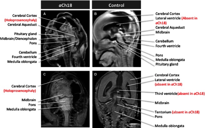
Fig. 2.
MRI phenotypic presentation of HPE in individual brain structures. (A) Sagittal view of HPE brain in aberrant Ch18 (aCh18), with features of anencephaly, absent anterior cranial fossa, and upright, enlarged brainstem structures. The control fetus (B) has a fully induced forebrain and well-developed lateral ventricle, which are both missing in aCh18. Coronal slice of aCh18 showing missing brain tissue on the right and lack of separation in the lobes (alobar). (D) The control for (C) exhibits the ventricles missing in aCh18, and the classic morphology of the brainstem.

