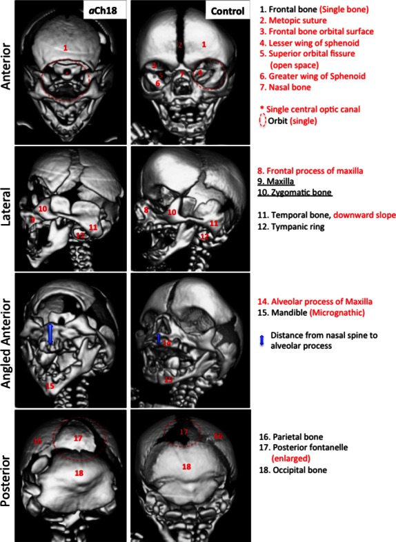
Fig. 6.

Craniofacial features of bone in aberrant Ch18 (aCh18) fetus compared with control. 3D reconstructed CT scans of a 28-week old aCh18 (left column) vs. a 29-week control fetus. Anterior (1st row), lateral (2nd row), angled anterior (3rd row) and posterior (4th row) perspectives of the cranium. Lateral features are preserved, whereas medial structures are severely impacted. Red lettering indicates an absence or abnormality in aCh18.
