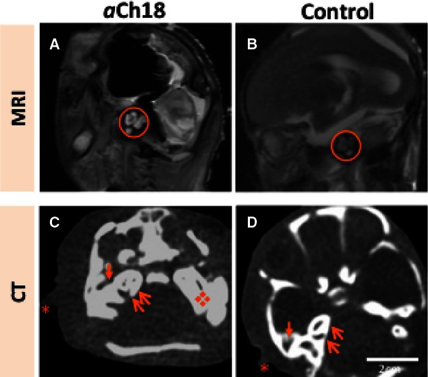
Fig. 7.

Internal, middle and external auditory structure development in HPE and cyclopia. (A) Sagittal magnetic resonance imaging (MRI) of the lateral head of an aberrant Ch18 (aCh18) fetus showing the middle auditory structures, mainly the ossicles (circle) appearing radiodense in aCh18 compared with (B) the age-matched control. (C) Horizontal section of the head at the level of the inner and external ear, and the petrous temporal bone showing their presence in radiodense form compared with (D) the age-matched control. Asterisk, pinna of ear; thin arrow, cochlea; thick arrow, external ear canal; diamond, enlarged petrous temporal bone. CT, computed tomography.
