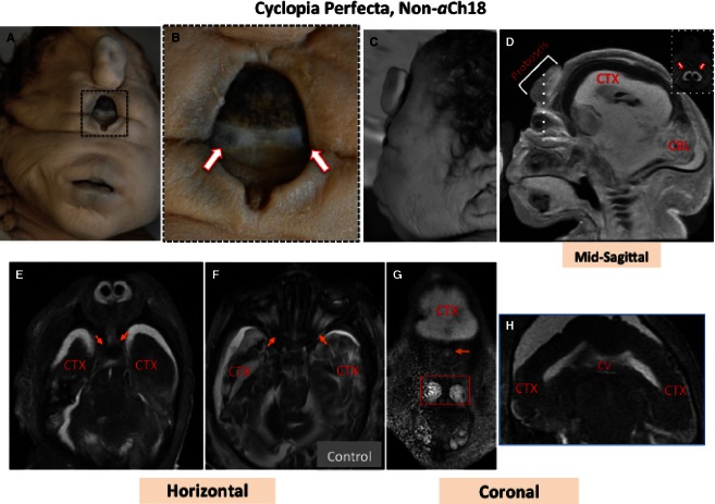
Fig. 8.
Superficial and MRI analysis of craniofacial features of a 35-week non-aberrant Ch18 (aCh18) cyclopic fetus with normal karyotype. (A, B) Frontal views of the 35-week cyclops with (B) enlargement of the eye to reveal two globes (arrows). (C) A lateral view of the head. (D) Sagittal, (E) horizontal, and (G and H) coronal sections through the head and face. (F) Age-matched control. The dashed line in (D) shows the plane of inset at the right. Arrows in (E) show bifurcating nerves as they enter the brain laterally. Arrows in (F) show nerves originating from two optic globes for normal binocular vision. The arrow in (G) shows a single optic nerve centrally, and the box reveals nasomaxillary structures. (H) A single cerebral ventricle (CV) in white connected at the midline, and unseparated cerebral cortex (CTX). CTX appears dark or light depending on if the images were, respectively, acquired with T1 or T2 weighting.

