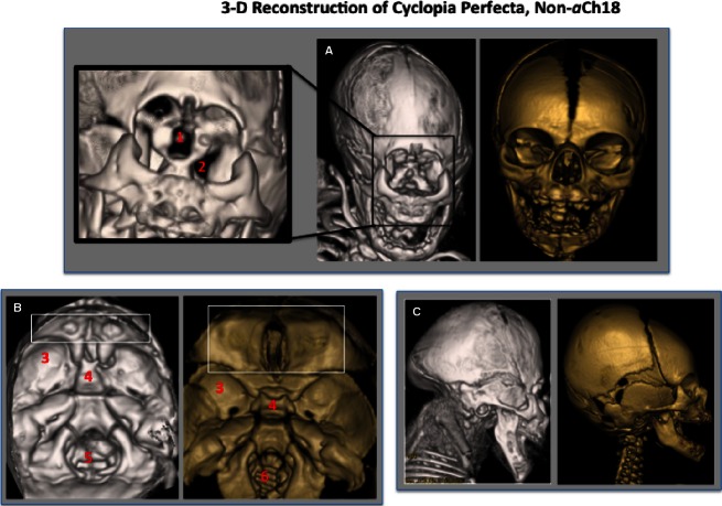
Fig. 9.
Analysis of the external cranium and internal floor of the cranium of a 35-week non-aberrant Ch18 (aCh18) cyclopia perfecta fetus in 3D. (A) Frontal view of the reconstructed face of cyclops (gray) with magnification at left showing defect in the orbit with a central slit for the optic nerve (1), but with lateral formation of a widened superior orbital fissure (2) compared with non-cyclopic control (gold). (B) Interior of the cranium with a small orbital plate of frontal bone with no crista galli (box) and anteriorly shifted middle cranial fossa (3), immature deviated sphenoid bone, and enlarged foramen magnum and petrous temporal bone. (C) Lateral aspect of the cranium, with unremarkable maxillary and mandibular structures, but with cranial defects in the temporal/occipital bones as well as abnormal cervical and thoracic vertebrae. Gray, 35-week cyclops; Gold, newborn control.

