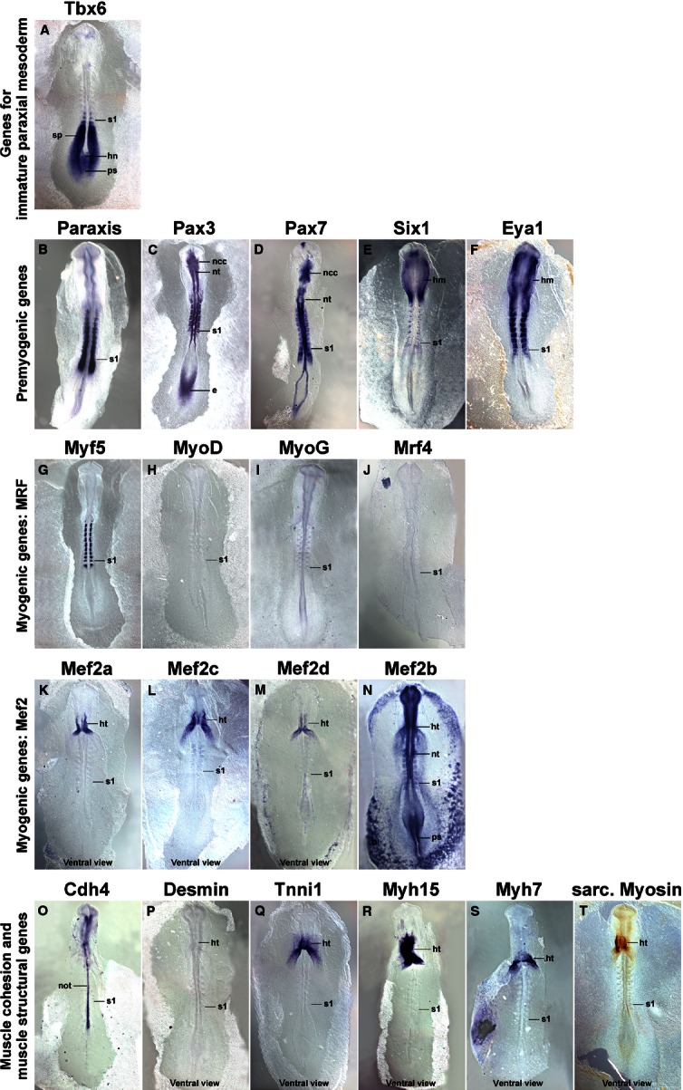
Fig. 2.

Marker gene expression at HH10. Dorsal views (P–T: ventral views), rostral to the top. Markers are indicated on top of each individual image as before. Tbx6 and the pre-myogenic genes show overlapping expression in the rostral segmental plate and the most recently formed somite. The pre-myogenic genes label the condensing as well as fully formed epithelial somites. Of the Mrf genes, Myf5 is expressed weakly in the condensing somite, and more robustly in the medial wall of the epithelial somites. Similar to HH8, Mef2c and 2d display some weak somitic expression, but the main expression of the Mef2 genes, of Tnni1 and the Myosins remains in heart (ht). Abbreviations see Fig. 1 and: bi, blood islands; ncc, neural crest cells; nt, neural tube; the position of the youngest somite is indicated (s1).
