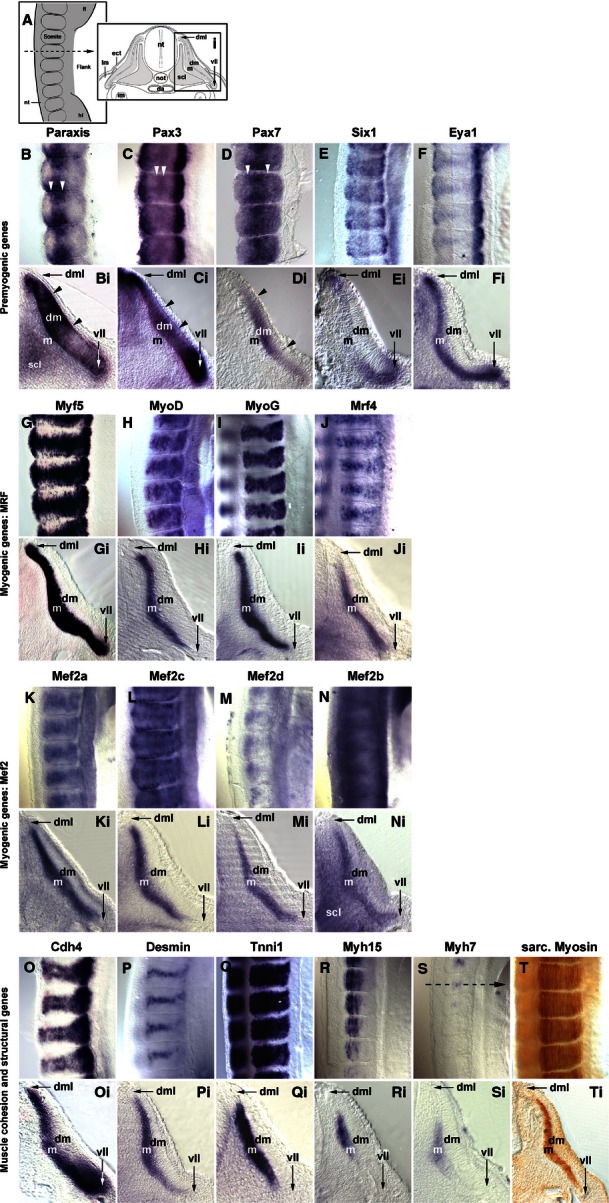
Fig. 5.

Marker gene expression in the flank of embryos at HH19-20. (A) Schematic representation of the images displayed in B–T (lateral view of flank somites on the right of the embryo, rostral to the top, lateral to the right) and Bi–Ti [cross section to flank somites, dorsal to the top, lateral to the right; section (Si) is from the forelimb- flank boundary as indicated in S]. Markers are indicated as before. Paraxis, Pax3 and Pax7 show distinct areas of elevated expression in the dermomyotome (B, Bi–D, Di; arrowheads). Their expression overlaps in dorsomedial and ventrolateral lips with that of Six1, Eya1 and Myf5; in the ventrolateral lip, expression overlaps also with that of Cdh4. The Mrf genes, the Mef2 genes and the genes encoding cell adhesion and muscle structural proteins show overlapping expression in the myotome, with the late commencing markers still being confined to the more medial territories. Abbreviations (see also Figs 1–3): da, dorsal aorta; dm, dermomyotome; dml, dorsomedial lip of dermomyotome; ect, surface ectoderm; fl, fore limb; hl, hind limb; m, myotome; scl, sclerotome; vll, ventrolateral lip of dermomyotome.
