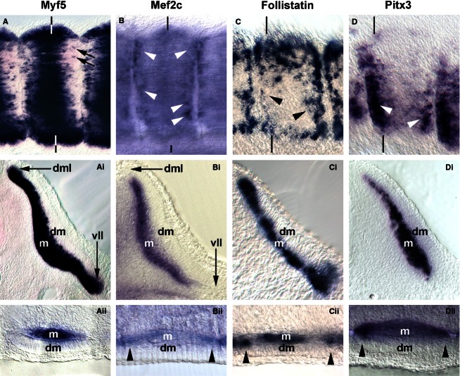
Fig. 8.
Comparison of markers labelling myogenic cells from the dorsomedial-ventrolateral and rostrocaudal lips of the dermomyotome. (A–D) Lateral views of flank somites on the right of the embryo, rostral to the right, dorsal to the top. (Ai–Di) Cross sections of these somites; (Ai, Bi) leading through the centre; (Ci,Di) sectioned along the caudal edge of the somite as indicated by the vertical lines. (Aii–Dii) Frontal sections, medial to the top, rostral to the right. Individual cells along the rostrocaudal sub-lip domain of the myotome express Myf5 (A, arrows). In contrast, robust and widespread expression in this domain is found for Mef2c, Follistatin and Pitx3 (B–D, Bii–Dii; arrowheads).

