
Figure 3. PKA inhibits α7 nAChR responses in HEK 293T cells.
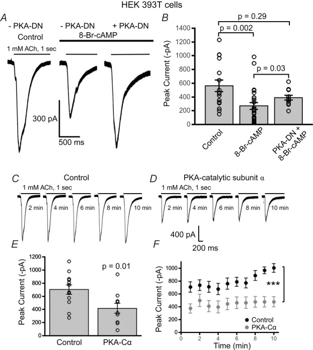
A, example waveforms of ACh (1 mm) evoked α7 nAChR responses from HEK 293T cells under three conditions including: control treatment, 8-Br-cAMP treatment and 8-Br-cAMP treatment with co-transfected dominant negative PKA (PKA-DN). B, 8-Br-cAMP (100 μm) (n = 26) stimulation resulted in a signficant attenuation of the mean α7 nAChR current as compared to control (n = 16) (P = 0.002, Wilcoxon rank sum test). Cells cotransfected with PKA-DN did not display a significant attenuation in nAChR current with 8-Br-cAMP as compared to cells only expressing α7 nAChRs (n = 11) (P = 0.29, Wilcoxon rank sum test). However, 8-Br-cAMP stimulation in α7-only transfected cells significantly attenuated α7 receptor currents as compared to cells co-transfected with PKA-DN (P = 0.03, Wilcoxon rank sum test). Sample traces of ACh (1 mm) evoked α7 nAChR responses from HEK 293T cells transiently expressing only α7 nAChRs (C) and α7 receptors co-expressed with PKA catalytic subunit α (PKA-Cα) (D). E, a significant decrease in the mean current amplitude of α7 nAChRs was observed in cells co-expressing PKA-Cα (n = 9) as compared to control (cells expressing only α7 nAChRs) (n = 12) (P = 0.01, t test). F, PKA-Cα and α7 nAChR co-transfected cells consistently showed reduced α7 nicotinic responses with repeated ACh applications as compared to control (α7 nAChRs only) (***P < 0.001, two-way ANOVA).