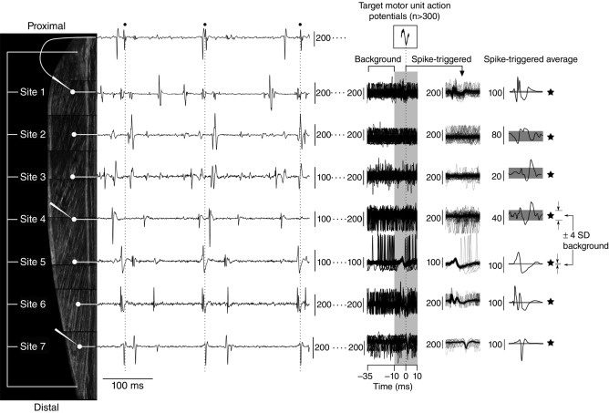
Figure 1. Experimental procedures and signal processing.
Seven multi-unit fine-wire electrodes (white circles) were inserted at even intervals along the medial gastrocnemius (MG) and three microelectrodes (white elongated triangles) were inserted as close as possible to the tips of the fine-wire electrodes located at Sites 1, 4 and 7. When subjects held a steady isometric contraction, the activity of several motor units (MUs) could be seen on the fine-wire recordings. At the same time the three microelectrodes recorded MU discharge activity (only signal from microelectrode at Site 1 included for clarity). The discharge times (black circles and dashed vertical lines) of selected MUs were used to spike-trigger each of the fine-wire recordings. In some instances it was possible to see potentials associated with the selected MU directly on the raw, overlayed multi-unit spike-triggered data (i.e. Sites 1, 5–7). In other instances the presence of an action potential associated with the selected MU was only visible in the spike-triggered average data (i.e. Sites 3 and 4). When a spike-triggered average had an action potential greater than four times the background signal (horizontal grey bands), it was considered that the selected MU had muscle fibres in close vicinity to the fine-wire recording electrode (black stars). Composite ultrasound image (subcutaneous tissue omitted) is from a subject with a 17 cm long MG muscle. The graphical location of fine-wire electrodes and microelectrodes was estimated from an insertion depth of 2 cm and an interelectrode distance of 2.5 cm. All vertical scale bars are in microvolts.

