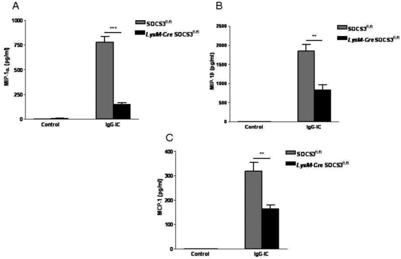
Figure 6.

Myeloid-specific disruption of SOCS3 resulted in decreased pro-inflammatory mediator expressions in IgG IC-treated lungs. SOCS3fl/fl and LysM-Cre SOCS3fl/fl mice were treated intratracheally with IgG IC for 4 h, and cell-free supernatants were used to conduct ELISA to detect MIP-1α (A), and MIP-1β (B), and MCP-1 (C) expressions. Values represented means ± S. E. M. for ≥ 3 mice for each group. *, and ** suggested statistically significant difference— p < 0.05, and p < 0.01, respectively.
